Welkom bij THIM Hogeschool voor Fysiotherapie & Bohn Stafleu van Loghum
THIM Hogeschool voor Fysiotherapie heeft ervoor gezorgd dat je Mijn BSL eenvoudig en snel kunt raadplegen. Je kunt je links eenvoudig registreren. Met deze gegevens kun je thuis, of waar ook ter wereld toegang krijgen tot Mijn BSL. Heb je een vraag, neem dan contact op met helpdesk@thim.nl.
Om ook buiten de locaties van THIM, thuis bijvoorbeeld, van Mijn BSL gebruik te kunnen maken, moet je jezelf eenmalig registreren. Dit kan alleen vanaf een computer op een van de locaties van THIM.
Eenmaal geregistreerd kun je thuis of waar ook ter wereld onbeperkt toegang krijgen tot Mijn BSL.
Login
Als u al geregistreerd bent, hoeft u alleen maar in te loggen om onbeperkt toegang te krijgen tot Mijn BSL.
A 21-year-old, previously healthy, female presented with complaints of palpitations. The physical examination, ECG and a 24-hour Holter investigation were unremarkable.
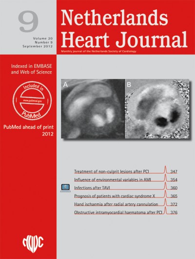
A transthoracic echocardiogram demonstrated a double orifice mitral valve (DOMV) (Fig. 1). Cardiac magnetic resonance imaging was performed in order to exclude other congenital heart problems (Fig. 2). There was no evidence of mitral valve stenosis, regurgitation or other cardiac morphological abnormalities.
Fig. 1
Transthoracic echocardiogram. a Apical 4-chamber view with ‘seagull’ appearance of the mitral valve (white arrow); b Parasternal short axis with bridge of tissue (dark arrow) dividing the mitral valve into two equal orifices
Fig. 2
Cardiac MRI. a Short-axis mitral valve gradient echo cine MRI image demonstrating the two orifices b) Corresponding phase contrast velocity encoded MRI (maximum velocity 100 cm/s) image demonstrating normal flow through the mitral valve
×
×
First described in 1876 by Greenfield [1], DOMV is an uncommon anomaly which, as its name indicates, has a single mitral annulus and opens into the left ventricle through two orifices. Depending on the relative size and location of the two orifices, DOMV can be classified into an eccentric type (found in 85% of cases) and a central or bridge type (as in our patient’s case) [2]. Mitral stenosis or regurgitation and other congenital malformations such as atrioventricular or ventricular septal defects may be associated with DOMV [3, 4].
Open Access
This article is distributed under the terms of the Creative Commons Attribution Noncommercial License which permits any noncommercial use, distribution, and reproduction in any medium, provided the original author(s) and source are credited.
Open AccessThis is an open access article distributed under the terms of the Creative Commons Attribution Noncommercial License (https://creativecommons.org/licenses/by-nc/2.0), which permits any noncommercial use, distribution, and reproduction in any medium, provided the original author(s) and source are credited.
Het Netherlands Heart Journal wordt uitgegeven in samenwerking met de Nederlandse Vereniging voor Cardiologie. Het tijdschrift is Engelstalig en wordt gratis beschikbaa ...