Welkom bij THIM Hogeschool voor Fysiotherapie & Bohn Stafleu van Loghum
THIM Hogeschool voor Fysiotherapie heeft ervoor gezorgd dat je Mijn BSL eenvoudig en snel kunt raadplegen. Je kunt je links eenvoudig registreren. Met deze gegevens kun je thuis, of waar ook ter wereld toegang krijgen tot Mijn BSL. Heb je een vraag, neem dan contact op met helpdesk@thim.nl.
Om ook buiten de locaties van THIM, thuis bijvoorbeeld, van Mijn BSL gebruik te kunnen maken, moet je jezelf eenmalig registreren. Dit kan alleen vanaf een computer op een van de locaties van THIM.
Eenmaal geregistreerd kun je thuis of waar ook ter wereld onbeperkt toegang krijgen tot Mijn BSL.
Login
Als u al geregistreerd bent, hoeft u alleen maar in te loggen om onbeperkt toegang te krijgen tot Mijn BSL.
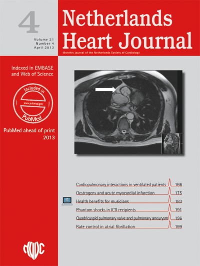
We report on a 37-year-old woman with essential systemic hypertension. An MRI was performed because of weak femoral pulses, depicting a severe almost atretic coarctation of the aorta (Fig. 1). Angiography of the proximal distal thoracic aorta was simultaneously performed (Fig. 2a). The transverse arch was narrow (16 mm), as was the diameter of the terminal aortic arch distal to the left subclavian artery (10 mm). Distal of the coarctation the diameter of the descending aorta was 11 mm. A trajectory of 2 mm in length seemed atretic. The atretic segment could be crossed in an antegrade fashion with a straight 0,014 in. coronary wire and balloon pre-dilatation was performed with a 5 mm coronary balloon. Thereafter, a multi-purpose catheter could be advanced retrogradely across the coarctation segment. A 9 French Mullins sheath (Cook) was advanced to the transverse aortic arch. A 41 mm long Advanta V12 premounted covered stent (Atrium, Hudson, USA) on a 12 mm high pressure balloon was implanted. Consecutive angiography revealed complete expansion of the stent up to 12 mm without residual stenosis, and no aneurysm formation (Fig. 2b).
Fig. 1
MRI imaging of the left ventricle (LV), ascending aorta (AAo). The left subclavian artery (LSA) is dilated and the descending aorta (DAo) is hypoplastic. The coarctation imposes as atresia. There are numerous collaterals. RV: right ventricle
Fig. 2
a Simultaneous angiography of the proximal and distal descending aorta; anterior projection. Atretic coarctation (CoA). Some small collaterals can be seen right of the coarctation. Abbreviations: Cath: catheter, DAo: descending aorta, LSA: left subclavian artery. L/R: left and right side of patient. b After stent-implantation (stent). Ao: transverse aorta. Left subclavian artery (LSA)
×
×
In conclusion, treatment with placement of a low-profile covered stent, using a simultaneous radial and femoral approach and pre-dilatation, delivered an excellent result without complications and a short hospital stay. The patient’s blood pressure returned to normal and her antihypertensive medication could be stopped within 3 weeks after stent implantation. We emphasise that in so-called ‘unexplained’ systemic hypertension, especially in young adults, coarctation of the aorta has to be excluded [1, 2]. When coarctation is confirmed primary stenting is the first choice therapeutic option [3‐5].
Open Access
This article is distributed under the terms of the Creative Commons Attribution Noncommercial License which permits any noncommercial use, distribution, and reproduction in any medium, provided the original author(s) and source are credited.
Open AccessThis is an open access article distributed under the terms of the Creative Commons Attribution Noncommercial License (https://creativecommons.org/licenses/by-nc/2.0), which permits any noncommercial use, distribution, and reproduction in any medium, provided the original author(s) and source are credited.
Het Netherlands Heart Journal wordt uitgegeven in samenwerking met de Nederlandse Vereniging voor Cardiologie. Het tijdschrift is Engelstalig en wordt gratis beschikbaa ...